
AC-26 - NuMA-like
Descrição
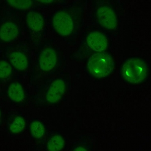
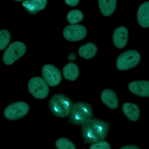
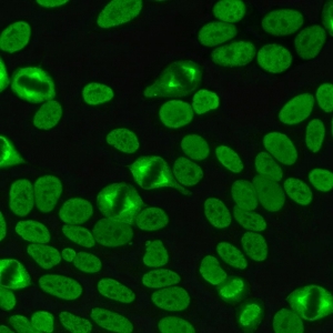
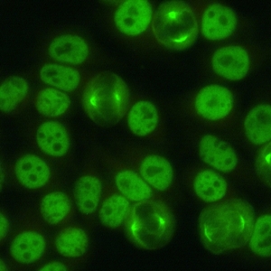
Nuclear speckled staining with spindle fibers.
Associação Antigênica
NuMA
Doença Associada
SjS, SLE, other
Relevância Clínica (Primeiro Nível)
Approximately one-half of the patients with the AC-26 pattern have clinical features of a SARD (SjS, SLE, UCTD, limited SSc, or RA); the AC-26 pattern is also observed in patients with organ-specific autoimmune diseases and less frequently in non-autoimmune conditions, especially when in low titer
Found very infrequently in a routine serology diagnostic setting
Antigens recognized include NuMA, centrophilin, SP-H antigen and NMP-22; specific immunoassays for these autoantibodies are currently not commercially available
Referências
Mozo L, Gutie?rrez C, Go?mez J, GA?3mez JesA?os. Antibodies to mitotic spindle apparatus: clinical significance of NuMA and HsEg5 autoantibodies. J Clin Immunol 2008;28:285–90.
Szalat R, Ghillani-Dalbin P, Jallouli M, et al. Anti-NuMA1 and anti-NuMA2 (anti- HsEg5) antibodies: clinical and Immunological features: a propos of 40 new cases and review of the literature. Autoimmun Rev 2010;9:652–6.
Grypiotis P, Ruffatti A, Tonello M, et al. [Clinical significance of fluoroscopic patterns specific for the mitotic spindle in patients with rheumatic diseases]. Reumatismo 2002;54:232–7.
Price CM, McCarty GA, Pettijohn DE. NuMA protein is a human autoantigen. Arthritis Rheum 1984;27:774–9.
Bonaci-Nikolic B, Andrejevic S, Bukilica M, et al. Autoantibodies to mitotic apparatus: association with other autoantibodies and their clinical significance. J Clin Immunol 2006;26:438–46.
Compton DA, Szilak I, Cleveland DW. Primary structure of NuMA, an intranuclear protein that defines a novel pathway for segregation of proteins at mitosis. J Cell Biol 1992;116:1395–408.